
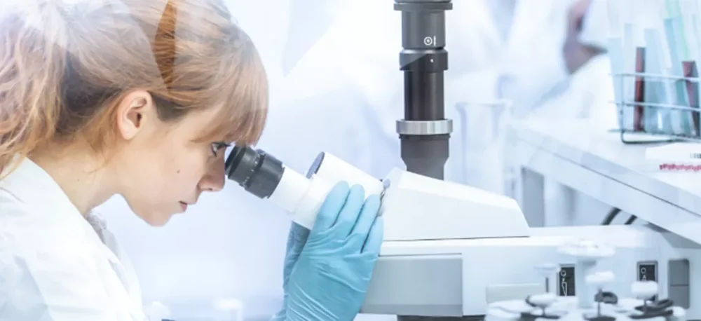
Data backed and driven solutions
All of our products have been tested and approved in both clinical and laboratory settings. We are constantly working with leading Healthcare Professionals to ensure that our solutions are meeting their needs, and the data speaks for itself. Check it out
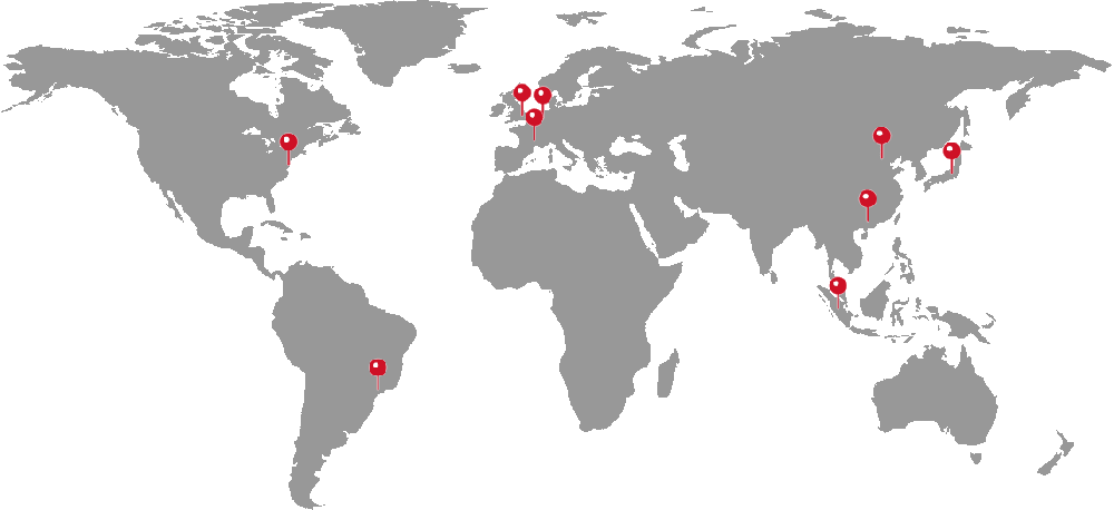
Global, Agile and Local
Since the inceptions of 3-D Matrix as a spinoff from MIT in 2004, 3-D Matrix has grown to become the leading global peptide solutions company, headquartered in Tokyo, Japan. With offices serving in America, Europe and Asia, our employees together with our partners and distributors serve research healthcare providers and patients around the world. Learn More


